Learn to differentiate between mast cell tumors and histiocytomas in dogs, including symptoms, diagnosis, and treatment options. Understand when to seek veterinary care.

This guide equips you to recognize key differences between mast cell tumors (MCTs) and histiocytomas, understand diagnostic methods, and know when to seek professional care. MCTs are cancerous, may spread, and require veterinary evaluation, while histiocytomas are benign and often resolve on their own. Both conditions demand accurate diagnosis for effective management.

This guide is for dog owners who notice lumps or growths on their pets and want to understand the differences between mast cell tumors and histiocytomas. It is intended for readers with basic knowledge of canine health but no veterinary training. This guide is not for those who have already received a confirmed diagnosis or are seeking immediate emergency care.
Distinguishing between mast cell tumors (MCTs) and histiocytomas is critical because MCTs are cancerous and can spread, while histiocytomas are benign and often resolve without intervention. This difference impacts treatment decisions and outcomes. Early detection ensures appropriate care, as MCTs require prompt action to prevent metastasis, whereas histiocytomas may not need treatment. Accurate diagnosis through veterinary evaluation is essential for effective management.
Before taking any action, be aware of the risks. If you notice any signs of systemic illness, such as vomiting, diarrhea, or severe swelling, stop and seek immediate veterinary care. Never attempt to diagnose or treat a lump without professional guidance, as misidentification can delay proper care.
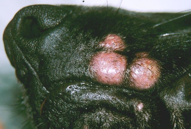
Histiocytomas are small, raised, red lumps that are typically found on the head, ears, or legs. These tumors are almost always circular and have a smooth surface. They are not itchy, which can help differentiate them from other types of skin growths. Histiocytomas are most common in dogs under 3 years of age and often resolve on their own over several weeks to months. They are benign and do not metastasize, meaning they do not spread to other parts of the body. While they may appear in short-haired dogs, they are not limited to this group. The exact cause of histiocytomas is not fully understood, but they are generally considered to be non-cancerous and have a good prognosis.
Mast cell tumors (MCTs) are cancerous growths that can appear in older dogs and may spread internally. These tumors can vary in appearance, ranging from benign-looking lumps to inflamed, irritated red growths or ulcers. One of the key symptoms of MCTs is itching, which can be persistent and uncomfortable for the dog. Swelling is also common, and in some cases, the tumor may cause systemic reactions such as vomiting, diarrhea, and stomach irritation. This is due to the release of histamines from the mast cells, which can affect the gastrointestinal tract. MCTs are more prevalent in certain breeds, including Boxers, Boston Terriers, Bulldogs, and Golden Retrievers. The severity of the condition depends on the tumor's grade, with low-grade tumors having a better prognosis than high-grade ones.
Fine needle aspiration (FNA) is a common and minimally invasive diagnostic method used to evaluate both histiocytomas and mast cell tumors (MCTs). During this procedure, a fine needle is inserted into the tumor to collect a sample of cells, which are then examined under a microscope. FNA can provide preliminary information about the nature of the growth, helping to distinguish between benign and malignant tumors. However, for a definitive diagnosis, histopathology is often required. This involves examining the tissue sample in a veterinary pathology laboratory to determine the exact type of tumor and its characteristics. Histopathology is crucial for confirming the diagnosis and guiding treatment decisions, especially for MCTs, where the tumor's grade and potential for metastasis play a significant role in determining the appropriate course of action.
Histiocytomas are most commonly found in dogs under 3 years of age, typically appearing as small, raised, red lumps on the head, ears, or legs. These tumors are generally benign and may resolve on their own without the need for treatment. In contrast, mast cell tumors (MCTs) are more prevalent in older dogs and certain breeds, including Boxers, Boston Terriers, and Golden Retrievers. The age and breed of the dog can influence the likelihood of developing these tumors. While histiocytomas are usually non-cancerous and have a good prognosis, MCTs are cancerous and can vary in aggressiveness. The breed-specific prevalence of MCTs highlights the importance of genetic factors in the development of these tumors, although the exact causes are not fully understood.
Histiocytomas often resolve on their own over several weeks to months, making them a relatively low-risk condition for dogs. In many cases, no treatment is necessary, and the tumor will disappear without intervention. However, some owners may choose to have the tumor removed for cosmetic reasons or to prevent potential complications. On the other hand, mast cell tumors (MCTs) typically require surgical removal, especially if they are cancerous or show signs of aggressiveness. Surgery is the primary treatment for MCTs, and the success of the procedure depends on the tumor's grade and the ability to remove it with wide margins. In some cases, additional treatments such as chemotherapy or radiation may be recommended, particularly for high-grade MCTs. The treatment approach for MCTs is more involved, and the prognosis can vary depending on the tumor's characteristics and the dog's overall health.
Surgical treatment for mast cell tumors (MCTs) requires wide margins to ensure complete removal of the tumor and reduce the risk of recurrence. The surgeon will remove the tumor along with a surrounding area of healthy tissue to minimize the chances of leaving behind any cancerous cells. This is particularly important for MCTs, as incomplete removal can lead to regrowth and a poorer prognosis. In aggressive cases, follow-up treatments such as radiation therapy or chemotherapy may be recommended. Radiation therapy is often used to target any remaining cancer cells after surgery, while chemotherapy may be prescribed to manage systemic spread or to shrink the tumor before surgery. The choice of treatment depends on the tumor's grade, location, and the dog's overall health, with the goal of achieving the best possible outcome.
Histiocytomas are benign tumors that do not metastasize, meaning they do not spread to other parts of the body. This makes them a less severe condition compared to mast cell tumors (MCTs), which are cancerous and can spread internally. MCTs are graded based on their risk of metastasis, with low-grade tumors having a better prognosis than high-grade ones. The grading system takes into account factors such as the tumor's size, location, and the presence of certain cellular characteristics. This information is crucial for determining the appropriate treatment plan and predicting the dog's outcome. For MCTs, a higher grade indicates a greater likelihood of spreading and a more aggressive treatment approach may be necessary. In contrast, histiocytomas generally have a favorable prognosis and do not require extensive treatment.
Both histiocytomas and mast cell tumors (MCTs) require veterinary consultation for accurate diagnosis and appropriate management. While these conditions can be visually similar, they have different implications for the dog's health and require distinct treatment approaches. A veterinarian can perform a thorough examination and use diagnostic methods such as fine needle aspiration or histopathology to determine the nature of the tumor. Early detection and proper diagnosis are crucial for ensuring the best possible outcome for the dog. Even though histiocytomas are generally benign and may resolve on their own, it is still important to consult a veterinarian to rule out more serious conditions. For MCTs, timely intervention is essential to prevent the tumor from spreading and to improve the chances of successful treatment.
Understanding the differences between mast cell tumors (MCTs) and histiocytomas is essential for effective diagnosis and treatment. While both conditions involve skin growths, they have distinct characteristics, risk factors, and treatment options. Histiocytomas are typically small, raised, red lumps that are benign and may resolve on their own, while MCTs are cancerous and can spread internally. Recognizing these differences helps pet owners and veterinarians make informed decisions about the best course of action. Early detection and proper management are key to ensuring the health and well-being of dogs affected by these tumors. By staying informed and seeking veterinary care when needed, pet owners can provide the best possible care for their pets.
Certain breeds, such as Boxers, Boston Terriers, and Golden Retrievers, are more prone to mast cell tumors (MCTs), which are cancerous and can spread. Histiocytomas, typically benign, are common in younger dogs regardless of breed. Understanding these variations helps tailor care based on your dog's risk factors and life stage.
Owners often mistake histiocytomas for MCTs or vice versa, leading to delayed treatment. Another mistake is attempting to treat a lump at home without professional guidance. Avoid these errors by seeking veterinary care for any new or changing lumps.
If your dog shows vomiting, diarrhea, or severe swelling, contact a veterinarian immediately. New or changing lumps, especially on the head, ears, or legs, require professional evaluation. These symptoms may indicate systemic illness or malignant growths.
Seek urgent care if lumps grow rapidly, bleed, or cause discomfort. Systemic signs like lethargy or loss of appetite also demand immediate attention.
This guide is informed by veterinary research and clinical observations, focusing on the differentiation between mast cell tumors and histiocytomas. It draws on established medical knowledge from sources such as veterinary pathologists and clinical studies to provide accurate and reliable information for dog owners.
If you notice any lumps on your dog, consult a veterinarian for proper diagnosis. Further information on diagnosis and treatment options is available through veterinary resources.
You might also like these articles.

Learn how desmopressin treats diabetes insipidus in dogs, including diagnosis, symptoms, and management strategies.

Learn to spot hidden cat illness symptoms and act early. Vets reveal key signs owners often miss, including appetite changes, weight shifts, and behavior shifts.